

- Updated: February 18, 2026
- 6 min read
MRI Study Finds 99% of Adults Over 40 Have Shoulder Abnormalities
Study Finds 99% of Adults Over 40 Show Rotator‑Cuff Abnormalities on MRI—What It Means for Your Shoulder Health
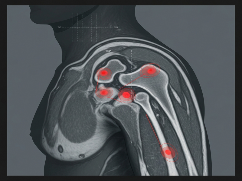
Answer: A new Finnish study published in JAMA Internal Medicine shows that 99 % of adults aged 40 and older have at least one rotator‑cuff abnormality on MRI, yet the overwhelming majority experience no shoulder pain or functional limitation.
Why This Study Matters
Shoulder pain is one of the most common musculoskeletal complaints worldwide, and magnetic resonance imaging (MRI) has become the go‑to diagnostic tool for clinicians trying to pinpoint the source. The recent Ars Technica report highlighted a startling statistic: 99 % of adults over 40 have “abnormalities” in their rotator cuff on MRI scans. This finding forces us to rethink how we interpret shoulder MRI results, especially when patients are asymptomatic.
Study Overview
Methodology & Sample Size
Finnish researchers leveraged a nationally representative health survey and invited participants to undergo bilateral shoulder MRI. A total of 602 volunteers aged 41‑76 completed the protocol, providing both self‑reported symptom data and high‑resolution imaging of each shoulder (1,204 shoulders in total). The study combined questionnaire responses with radiologist‑graded MRI findings, allowing a direct comparison between structural changes and reported pain.
Key Statistics
- 99 % (595/602) of participants displayed at least one rotator‑cuff abnormality.
- Partial‑thickness tears were the most common (62 %).
- Tendinopathy (tendon swelling) appeared in 25 % of scans.
- Full‑thickness tears were present in 11 % of participants, increasing sharply after age 70.
- 82 % (492/602) reported no shoulder symptoms, yet 96 % of their asymptomatic shoulders still showed abnormalities.
- Only 18 % (110/602) reported pain, and 98 % of those painful shoulders also had MRI findings, but the prevalence of partial tears and tendinopathy was nearly identical to the asymptomatic group.
Age‑Related Progression
The data revealed a clear age gradient: participants under 45 showed virtually no full‑thickness tears, while those aged 70‑76 exhibited the highest rates. This suggests that many rotator‑cuff changes are part of normal aging rather than pathological lesions demanding surgical repair.
Clinical Implications: When MRI May Mislead
The study’s headline‑grabbing statistic can be misinterpreted as a call for more aggressive imaging, but the opposite is true. Below are the practical takeaways for clinicians and patients alike:
- Incidental findings are the norm. An MRI “abnormality” in a 55‑year‑old is more likely a benign age‑related change than a surgically correctable tear.
- Symptom correlation matters more than imaging. Treatment decisions should prioritize pain severity, functional limitation, and physical‑exam findings over the mere presence of a tear.
- Over‑use of MRI drives overtreatment. Unnecessary scans increase healthcare costs and may lead patients to pursue invasive procedures that offer little benefit.
- Terminology influences perception. Describing findings as “lesions” or “degeneration” rather than “tears” can reduce patient anxiety and the perceived need for immediate repair.
In the era of AI‑enhanced diagnostics, platforms like the Enterprise AI platform by UBOS can help clinicians triage imaging results by integrating symptom scores, activity data, and predictive models—ensuring that MRI findings are interpreted in a broader clinical context.
Expert Commentary: Imaging Alone Is Not Enough
Orthopedic surgeons at the University of California, San Francisco, emphasized that “MRI should be a tool, not a verdict.” They argue that the language used in radiology reports often overstates the clinical relevance of age‑related changes. Dr. Edgar Garcia‑Lopez noted:
“When we label a partial‑thickness tear as a ‘tear,’ patients assume it must be fixed. In reality, many of these findings are incidental and do not limit function.”
The authors of the study also recommend a shift toward more precise descriptors such as “fraying” or “structural alteration.” This nuanced language aligns with the emerging AI marketing agents approach of personalizing communication—only now applied to medical reporting.
What This Means for You: Actionable Shoulder‑Health Tips
If you’re over 40 and concerned about shoulder pain, consider the following evidence‑based steps before requesting an MRI:
- Start with a functional assessment. A qualified physical therapist can evaluate range of motion, strength, and pain patterns without imaging.
- Try a structured exercise program. Strengthening the rotator cuff and scapular stabilizers reduces pain for most non‑traumatic cases. The over‑40 exercise tips page offers a free routine.
- Use a trial of conservative care. Two to three months of targeted physiotherapy, activity modification, and anti‑inflammatory measures often resolves symptoms.
- Reserve MRI for red‑flag cases. Persistent pain, night‑time discomfort, or a clear history of trauma may justify imaging.
- Interpret MRI results with a specialist. Ensure the radiologist’s report is reviewed alongside your clinical exam; ask for language that reflects “degeneration” rather than “tear” when appropriate.
- Leverage AI‑driven health tools. Apps built on the Web app editor on UBOS can track shoulder‑specific metrics, flagging when a professional evaluation is truly needed.
For those interested in integrating AI into personal health monitoring, the Workflow automation studio lets you create custom alerts—e.g., “Notify me if shoulder pain persists > 4 weeks.” This proactive approach reduces unnecessary imaging while ensuring timely care.
Bottom Line
The Finnish rotator‑cuff study underscores a simple truth: most shoulder “abnormalities” seen on MRI in adults over 40 are benign and asymptomatic. Clinicians should prioritize patient history, functional testing, and conservative management before ordering advanced imaging. Patients, meanwhile, can adopt evidence‑based exercise, seek professional assessment, and use AI‑enabled tools to stay informed without over‑relying on scans.
By aligning expectations with the reality of age‑related shoulder changes, we can curb MRI overuse, avoid unnecessary surgeries, and focus on interventions that truly improve quality of life.
Read the full study coverage on Ars Technica.
For more on how AI can streamline health data, explore the AI Video Generator or the AI SEO Analyzer to see how intelligent platforms improve decision‑making. If you’re a startup looking to build health‑focused applications, the UBOS for startups program offers a fast‑track to market. Enterprises can also benefit from the UBOS partner program, which includes dedicated support for integrating medical imaging workflows.
Need a quick prototype? Check out the UBOS templates for quick start, or dive into the UBOS pricing plans to find a tier that fits your practice’s budget.
Finally, for developers interested in conversational health assistants, the ChatGPT and Telegram integration and the OpenAI ChatGPT integration provide a foundation for building AI‑driven patient triage bots.
Andrii Bidochko
CTO UBOS
Andrii Bidochko is an AI entrepreneur and researcher focused on AI agents, reinforcement learning, and autonomous systems. He writes about the technologies shaping the future of machine intelligence, from frontier models and agent architectures to real-world AI applications.