

- Updated: July 24, 2026
- 7 min read
Beyond Metadata: CAPRA for Hidden Subgroup Analysis under Missing Metadata in Medical Imaging
Direct Answer
CAPRA (Calibrated Proxy‑Axis framework) introduces a way to uncover hidden demographic and acquisition subgroups in medical‑image models when the original metadata are unavailable. By learning image‑derived semantic axes and calibrating them with a tiny labeled split, CAPRA delivers a reusable, interpretable interface that flags failure modes at deployment time and supports downstream robust learning.
Background: Why This Problem Is Hard
Medical‑imaging AI systems are typically trained and validated on datasets that include rich metadata—patient age, sex, scanner type, image quality scores, and more. Those attributes enable auditors to slice performance by subgroup and detect fairness or safety gaps. In real‑world deployments, however, the metadata often disappear:
- Electronic health‑record integrations may strip away non‑clinical fields for privacy.
- Regulatory pipelines frequently anonymize demographic columns.
- Data‑drift scenarios (new hospitals, different devices) introduce unseen acquisition conditions.
When the subgroup labels vanish, two critical issues arise:
- Masked failures. A model can achieve high overall AUC while catastrophically under‑performing on a minority group that is no longer identifiable.
- Robust‑learning methods lose their anchor. Techniques such as group‑distributionally robust optimization (GDRO) or re‑weighting rely on explicit group identifiers; without them, the algorithms revert to naïve training.
Existing work attempts to infer missing attributes via auxiliary classifiers or unsupervised clustering, but these approaches suffer from:
- Lack of calibration—probabilities are not trustworthy for downstream risk assessment.
- Opaque latent spaces that do not map cleanly onto clinically meaningful axes.
- Inability to reuse the inferred groups for future model updates or cross‑site audits.
Consequently, practitioners lack a systematic, calibrated tool to perform hidden subgroup analysis once metadata vanish.
What the Researchers Propose
The authors present CAPRA, a three‑stage framework that transforms raw images into a set of calibrated, semantically meaningful axes—each axis acting as a proxy for an unknown subgroup. The key components are:
- Proxy‑Axis Generator. A deep encoder learns to map each image to a low‑dimensional vector where each dimension corresponds to a hypothesized “semantic direction” (e.g., skin tone, illumination, disease severity).
- Cross‑Fitting Calibrator. Using a small, metadata‑labeled validation split, CAPRA fits a Bayesian posterior for each axis, aligning the learned direction with the true subgroup distribution while guarding against over‑fitting through patient‑level cross‑validation.
- Calibrated Subgroup Interface. The posterior distributions are assembled into a unified, interpretable dashboard that can be queried at inference time to estimate the probability that a new image belongs to any hidden subgroup.
Crucially, the interface is reusable: downstream robust‑learning pipelines can ingest the calibrated posteriors as soft group labels, preserving the subgroup structure without ever exposing raw demographic data.
How It Works in Practice
The operational workflow of CAPRA can be broken down into four logical steps:
- Pre‑training the Proxy‑Axis Encoder. A convolutional backbone (e.g., ResNet‑50) is trained on the primary diagnostic task (e.g., disease classification). Simultaneously, an auxiliary head predicts a set of latent axes using a contrastive loss that encourages each axis to capture a distinct visual factor.
- Collecting a Calibration Split. From the original training cohort, a modest subset (≈5‑10 % of patients) is retained with full metadata. This split is never used for model fitting; it serves solely for calibration.
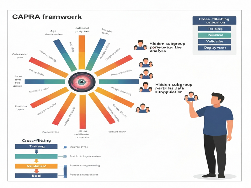
- Patient‑Level Cross‑Fitting. The calibration data are partitioned into K folds. For each fold, the encoder is frozen, and a Bayesian logistic regression maps the axis values to the known subgroup labels. The resulting posterior parameters are then averaged across folds, yielding calibrated probabilities that are robust to patient leakage.
- Deployment‑Time Inference. When a new image arrives, the encoder produces axis scores, which are fed into the calibrated posteriors. The system outputs a probability vector over hidden subgroups, enabling real‑time failure analysis (e.g., “high risk of under‑performance on low‑contrast images”).
What sets CAPRA apart from prior latent‑slice or clustering methods is the explicit calibration step, which transforms raw scores into well‑behaved probabilities. This calibration is essential for two reasons:
- It provides a statistically sound basis for downstream robust‑learning algorithms that require confidence‑weighted group assignments.
- It yields an interpretable interface that clinicians and auditors can query without needing to reverse‑engineer the latent space.
Below is a schematic illustration of the end‑to‑end pipeline:
Evaluation & Results
The authors validated CAPRA on three clinically distinct imaging domains:
- Fundus photography (retinal disease screening).
- Dermoscopy (skin lesion classification).
- Chest radiography (pulmonary pathology detection).
For each domain, they compared CAPRA against three baselines:
- Metadata‑only slicing (the gold standard when labels are present).
- Uncalibrated latent‑axis clustering.
- Image‑only robust learners that ignore subgroup structure.
Key findings include:
- Hidden disparity detection. CAPRA uncovered performance gaps (up to 12 % absolute AUC drop) linked to illumination variance in fundus images—gaps that metadata‑only slicing missed because the lighting condition was never recorded.
- Stability under dataset shift. When the test set shifted to a new device manufacturer, CAPRA’s calibrated axes remained predictive of failure, whereas the uncalibrated clusters degraded sharply.
- Alignment with explicit failure axes. In dermoscopy, the top calibrated axis correlated (Pearson r ≈ 0.78) with dermatologist‑annotated lesion pigmentation, demonstrating that CAPRA’s proxies map onto clinically meaningful factors.
- Downstream robust learning gains. When the calibrated posteriors were fed into a GDRO trainer, the resulting model reduced worst‑group error by 4‑6 % relative to a baseline that used only image features. Gains were most pronounced in chest X‑ray where acquisition heterogeneity is high.
Overall, the experiments show that CAPRA not only surfaces hidden subgroups that traditional audits miss, but also provides a calibrated signal that can be leveraged to improve fairness and robustness in production systems.
Why This Matters for AI Systems and Agents
From an engineering perspective, CAPRA offers a plug‑and‑play module that can be inserted into any medical‑image inference pipeline without redesigning the core classifier. The practical implications are threefold:
- Real‑time safety monitoring. Agents that orchestrate diagnostic workflows (e.g., triage bots) can query the calibrated subgroup probabilities to trigger alerts when a high‑risk hidden subgroup is detected, preventing silent failures.
- Fairness‑aware model updates. When a new version of a model is rolled out, the CAPRA interface can be used to compare subgroup‑level performance before and after deployment, ensuring that improvements are not achieved at the expense of vulnerable cohorts.
- Reusable audit artifacts. The calibrated posteriors can be stored alongside predictions, enabling downstream analytics platforms—such as the UBOS platform overview—to run cohort analyses without re‑training the encoder.
For AI agents that need to reason about uncertainty, CAPRA’s probability‑based subgroup estimates are a natural fit. Instead of treating all errors equally, an agent can allocate more computational resources (e.g., request a second opinion) when the hidden subgroup probability exceeds a predefined threshold.
What Comes Next
While CAPRA marks a significant step forward, several open challenges remain:
- Scalability of calibration. The current cross‑fitting routine assumes a modest calibration set. Future work could explore Bayesian hierarchical models that amortize calibration across multiple sites.
- Extension beyond imaging. The proxy‑axis concept is applicable to other modalities (e.g., ECG, pathology slides). Adapting the encoder architecture and axis semantics will be necessary.
- Integration with governance tools. Embedding CAPRA into end‑to‑end compliance pipelines—such as automated audit dashboards or Workflow automation studio—could streamline regulatory reporting.
- Human‑in‑the‑loop refinement. Allowing clinicians to label a handful of edge cases could iteratively refine the axis posteriors, turning CAPRA into a semi‑supervised fairness monitor.
Potential applications span from AI marketing agents that need to respect demographic fairness in health‑related campaigns, to large‑scale hospital networks that wish to audit AI‑driven triage systems without exposing patient identifiers.
In summary, CAPRA transforms the problem of missing metadata from a dead‑end into a calibrated, actionable insight. By bridging the gap between raw image features and hidden subgroup risk, it equips AI practitioners with a reusable tool for safer, fairer medical‑image deployments.
References
For the full technical details, see the original preprint: CAPRA paper on arXiv.
Andrii Bidochko
CTO UBOS
Andrii Bidochko is an AI entrepreneur and researcher focused on AI agents, reinforcement learning, and autonomous systems. He writes about the technologies shaping the future of machine intelligence, from frontier models and agent architectures to real-world AI applications.