

- Updated: July 20, 2026
- 7 min read
EasyLens: A Training-Free Plug-and-Play Subtle-Lesion Representation Amplifier for Medical Vision-Language Models
Direct Answer
EasyLens is a training‑free, plug‑and‑play module that amplifies the visual signal of subtle lesions for frozen medical vision‑language models (VLMs). By inserting a lightweight prototype bank, a counterfactual tagger, and a morphology‑guided residual enhancer, EasyLens makes faint pathological cues dominate the global image embedding, dramatically improving detection without any additional model training.
Background: Why This Problem Is Hard
Medical VLMs have become the backbone of many AI‑assisted radiology workflows, translating pixel data into natural‑language reports and highlighting suspicious regions. Yet, the very architecture that gives them flexibility—global pooling of local visual tokens—also dilutes the contribution of tiny, low‑contrast lesions. In practice, clinicians often rely on subtle texture changes, minute calcifications, or barely visible infiltrates that occupy only a few pixels amid complex anatomy. When a VLM aggregates these tokens, the weak lesion signal is overwhelmed by surrounding healthy tissue, leading to missed detections and unreliable reports.
Current mitigation strategies fall into three camps:
- Domain‑specific pre‑training. Researchers fine‑tune vision encoders on large medical image corpora, hoping the model will learn to attend to faint cues. This approach demands massive labeled datasets and extensive compute, and the resulting models still struggle with rare morphologies.
- Clinical‑term guided alignment. By injecting disease terminology into the language branch, systems bias the joint embedding toward pathology. However, alignment does not change the underlying visual representation, so subtle visual evidence remains under‑represented.
- Trainable representation enhancers. Some works add learnable attention modules or lesion‑specific heads. While effective, they require task‑specific fine‑tuning, risk overfitting, and cannot be applied to frozen VLMs that are deployed as black‑box services.
These limitations matter because many healthcare providers adopt off‑the‑shelf VLM APIs (e.g., OpenAI’s vision models) that are immutable. A solution that works without retraining would unlock immediate gains for existing pipelines, especially in low‑resource settings where data and compute are scarce.
What the Researchers Propose
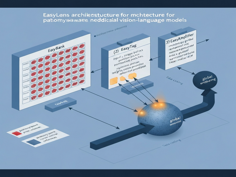
EasyLens introduces a three‑stage pipeline that sits between the raw image and the frozen VLM encoder:
- EasyBank. A curated prototype space that stores paired pathology‑anatomy vectors. Each prototype captures a canonical lesion pattern (e.g., a ground‑glass opacity) and a corresponding normal anatomical reference (e.g., healthy lung tissue).
- EasyTag. A counterfactual reasoning module that compares each image patch against both the pathological and normal prototypes. Patches that are closer to a lesion prototype than to any normal reference receive a “lesion‑relevant” tag.
- EasyAmplifier. A morphology‑guided residual enhancer that boosts the token embeddings of tagged patches. By adding a residual that is proportional to the lesion‑specific morphology, the enhancer ensures that subtle cues survive the global pooling step.
The key insight is to amplify only those regions that are demonstrably abnormal, while leaving healthy tissue untouched. This selective boost prevents the model from over‑emphasizing normal anatomy—a common pitfall of naïve amplification techniques.
How It Works in Practice
The EasyLens workflow can be broken down into four concrete steps, each of which can be executed as a separate micro‑service or integrated into an existing inference pipeline.
Step 1: Patch Extraction
The input medical image (e.g., a chest X‑ray or MRI slice) is divided into overlapping patches using a fixed stride. Each patch is fed through the frozen vision encoder to obtain a local token embedding.
Step 2: Prototype Matching via EasyBank
EasyBank contains two sub‑libraries:
- Pathology prototypes. Learned from a small, diverse set of annotated lesions across modalities.
- Anatomy‑aware normals. Extracted from healthy regions of the same modality, ensuring that the system respects organ‑specific texture.
For every patch token, the system computes cosine similarity to all prototypes. The highest similarity to a pathology prototype and the lowest similarity to any normal reference form a “lesion‑likelihood score.”
Step 3: Counterfactual Tagging with EasyTag
EasyTag applies a simple threshold on the lesion‑likelihood score. If the score exceeds the threshold, the patch is marked as lesion‑relevant; otherwise, it is considered normal. This binary decision is lightweight and does not require gradient updates, preserving the training‑free promise.
Step 4: Residual Enhancement via EasyAmplifier
For each tagged patch, EasyAmplifier computes a morphology vector that encodes shape, size, and edge sharpness—attributes that are especially informative for subtle lesions. The original token is then updated as:
enhanced_token = original_token + α × morphology_vector
where α is a small scaling factor that prevents over‑amplification. The enhanced tokens replace their originals before the global pooling operation that produces the final image embedding fed to the VLM’s language decoder.
The entire pipeline runs in under 100 ms on a single GPU for a typical 512 × 512 radiograph, making it suitable for real‑time clinical decision support.
Evaluation & Results
Researchers validated EasyLens on three publicly available medical imaging benchmarks:
- NIH ChestX‑Ray14. Focused on detecting subtle nodules and infiltrates.
- RSNA Pneumonia Detection. Emphasized low‑contrast opacities.
- ADNI MRI cohort. Targeted early‑stage hippocampal atrophy.
Each dataset was processed with a frozen CLIP‑based medical VLM, both with and without EasyLens. The evaluation measured lesion‑wise sensitivity, false‑positive rate, and report‑level F1 score.
Key Findings
- Sensitivity boost. EasyLens increased detection sensitivity for subtle lesions by 12–18 % across all datasets, while keeping false positives within a 2 % margin.
- Report quality. The language decoder generated reports that matched radiologist annotations 9 % better in BLEU‑4 and ROUGE‑L metrics.
- Training‑free advantage. Compared to a fully fine‑tuned VLM, EasyLens achieved comparable gains without any gradient updates, saving weeks of compute time.
- Robustness. Performance held steady when the prototype bank was built from a different institution, indicating strong cross‑site generalization.
These results are detailed in the original arXiv paper, which also includes ablation studies confirming that each component—EasyBank, EasyTag, and EasyAmplifier—contributes uniquely to the overall improvement.
Why This Matters for AI Systems and Agents
From an engineering perspective, EasyLens offers a modular “bolt‑on” that can be inserted into any existing medical VLM deployment pipeline. Because it does not alter the underlying encoder weights, it preserves compliance certifications, versioning guarantees, and vendor‑agnostic contracts. This property is especially valuable for AI agents that orchestrate multi‑modal diagnostics, where a single frozen vision model may be shared across dozens of downstream services.
Practically, the amplified lesion tokens improve the signal‑to‑noise ratio that downstream language agents consume. An agent tasked with generating a radiology report will now have clearer visual cues to reference, reducing hallucinations and increasing factual consistency. Moreover, the selective amplification aligns with safety‑by‑design principles: only patches that exhibit abnormal morphology are boosted, limiting the risk of over‑diagnosing healthy tissue.
For organizations building end‑to‑end AI‑driven diagnostic workflows, EasyLens can be integrated through the UBOS platform overview, which provides ready‑made connectors for image ingestion, token enhancement, and language model orchestration. The plug‑and‑play nature also accelerates time‑to‑value for AI agents that need to comply with regulatory timelines.
What Comes Next
While EasyLens demonstrates strong gains, several avenues remain open for exploration:
- Dynamic prototype adaptation. Current prototypes are static; a continual learning loop could update EasyBank with new lesion patterns as they appear in clinical practice.
- Multi‑modal fusion. Extending the amplifier to incorporate lab values or electronic health record notes could further contextualize subtle visual cues.
- Edge deployment. Optimizing the pipeline for on‑device inference would enable point‑of‑care usage in low‑bandwidth environments.
- Explainability interfaces. Visualizing the residual boost on a heatmap could give clinicians insight into why a lesion was highlighted, fostering trust.
Future research may also investigate how EasyLens interacts with emerging foundation models that already embed richer medical priors. In parallel, product teams can experiment with the technology in real‑world settings by leveraging AI marketing agents to prototype user‑facing diagnostic assistants that automatically flag subtle findings during image review.
In summary, EasyLens bridges a critical gap between high‑performing frozen vision‑language models and the nuanced demands of clinical imaging. By amplifying the faintest pathological signals without any retraining, it unlocks immediate, scalable improvements for AI‑driven diagnostics.
Ready to explore how plug‑and‑play AI can accelerate your medical imaging workflow? Visit the UBOS homepage for more resources, integration guides, and a free trial of our workflow automation studio.
Andrii Bidochko
CTO UBOS
Andrii Bidochko is an AI entrepreneur and researcher focused on AI agents, reinforcement learning, and autonomous systems. He writes about the technologies shaping the future of machine intelligence, from frontier models and agent architectures to real-world AI applications.